 Movie
Movie Controller


+ Open data

- Basic information
| Entry | Database: EMDB / ID: EMD-1757 | |||||||||
|---|---|---|---|---|---|---|---|---|---|---|
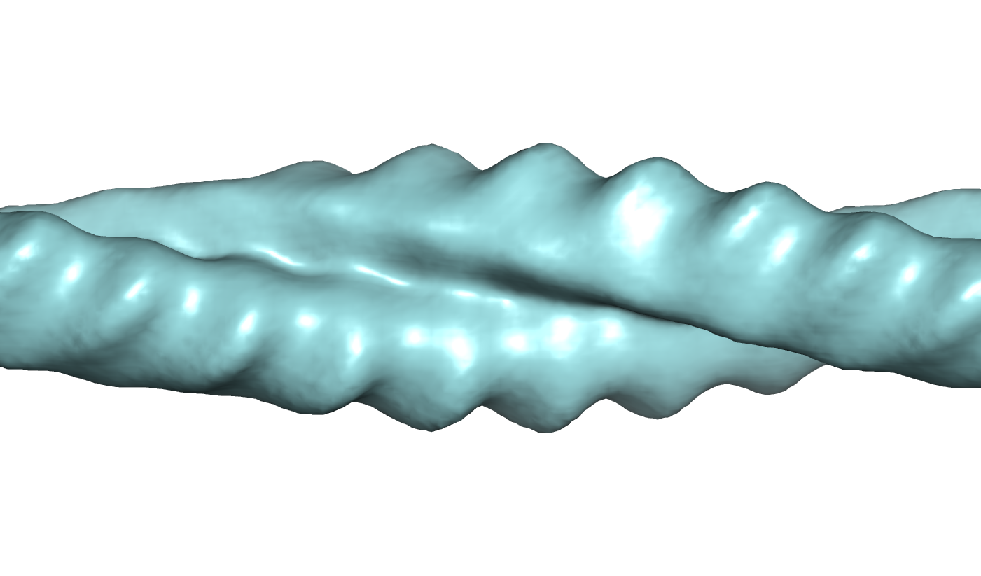
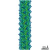
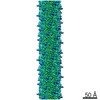
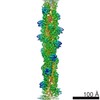
| Title | The Structure of TubZ filaments | |||||||||
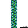
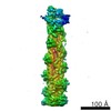
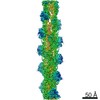
 Map data Map data | Double helical filaments of TubZ (pBT156) (Uniprot Q8KNP3) from Bacillus thuringiensis serovar israelensis (ATCC 35646) - negatively stained | |||||||||
 Sample Sample |
| |||||||||
 Keywords Keywords |  Cytoskeletal / DNA segregation / FtsZ / FtsZ-like / pBtoxis / pBT156 / plasmid partitioning / RepX / tubulin / tubulin-like / TubZ Cytoskeletal / DNA segregation / FtsZ / FtsZ-like / pBtoxis / pBT156 / plasmid partitioning / RepX / tubulin / tubulin-like / TubZ | |||||||||
| Biological species |  Bacillus thuringiensis (bacteria) Bacillus thuringiensis (bacteria) | |||||||||
| Method | helical reconstruction / negative staining / Resolution: 35.0 Å | |||||||||
 Authors Authors | Aylett CHS / Amos LA / Lowe J | |||||||||
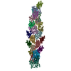
 Citation Citation | Journal: Proc Natl Acad Sci U S A / Year: 2010 Title: Filament structure of bacterial tubulin homologue TubZ. Authors: Christopher H S Aylett / Qing Wang / Katharine A Michie / Linda A Amos / Jan Löwe /  Abstract: Low copy number plasmids often depend on accurate partitioning systems for their continued survival. Generally, such systems consist of a centromere-like region of DNA, a DNA-binding adaptor, and a ...Low copy number plasmids often depend on accurate partitioning systems for their continued survival. Generally, such systems consist of a centromere-like region of DNA, a DNA-binding adaptor, and a polymerizing cytomotive filament. Together these components drive newly replicated plasmids to opposite ends of the dividing cell. The Bacillus thuringiensis plasmid pBToxis relies on a filament of the tubulin/FtsZ-like protein TubZ for its segregation. By combining crystallography and electron microscopy, we have determined the structure of this filament. We explain how GTP hydrolysis weakens the subunit-subunit contact and also shed light on the partitioning of the plasmid-adaptor complex. The double helical superstructure of TubZ filaments is unusual for tubulin-like proteins. Filaments of ParM, the actin-like partitioning protein, are also double helical. We suggest that convergent evolution shapes these different types of cytomotive filaments toward a general mechanism for plasmid separation. | |||||||||
| History |
|
- Structure visualization
| Movie |
 Movie viewer Movie viewer |
|---|---|
| Structure viewer | EM map: SurfViewMolmilJmol/JSmol |
| Supplemental images |
- Downloads & links
-EMDB archive
| Map data | emd_1757.map.gz | 67.8 KB | EMDB map data format | |
|---|---|---|---|---|
| Header (meta data) | emd-1757-v30.xmlemd-1757.xml | 8.4 KB 8.4 KB | Display Display | EMDB header |
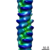
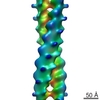
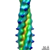
| Images |  1757.png 1757.png | 341.1 KB | ||
| Archive directory |  http://ftp.pdbj.org/pub/emdb/structures/EMD-1757ftp://ftp.pdbj.org/pub/emdb/structures/EMD-1757 http://ftp.pdbj.org/pub/emdb/structures/EMD-1757ftp://ftp.pdbj.org/pub/emdb/structures/EMD-1757 | HTTPS FTP |
-Related structure data
-Links
| EMDB pages | EMDB (EBI/PDBe) / EMDataResource |
|---|
-Map
| File | Download / File: emd_1757.map.gz / Format: CCP4 / Size: 666 KB / Type: IMAGE STORED AS FLOATING POINT NUMBER (4 BYTES) | ||||||||||||||||||||||||||||||||||||||||||||||||||||||||||||||||||||
|---|---|---|---|---|---|---|---|---|---|---|---|---|---|---|---|---|---|---|---|---|---|---|---|---|---|---|---|---|---|---|---|---|---|---|---|---|---|---|---|---|---|---|---|---|---|---|---|---|---|---|---|---|---|---|---|---|---|---|---|---|---|---|---|---|---|---|---|---|---|
| Annotation | Double helical filaments of TubZ (pBT156) (Uniprot Q8KNP3) from Bacillus thuringiensis serovar israelensis (ATCC 35646) - negatively stained | ||||||||||||||||||||||||||||||||||||||||||||||||||||||||||||||||||||
| Projections & slices | Image control
Images are generated by Spider. generated in cubic-lattice coordinate | ||||||||||||||||||||||||||||||||||||||||||||||||||||||||||||||||||||
| Voxel size | X=Y=Z: 4 Å | ||||||||||||||||||||||||||||||||||||||||||||||||||||||||||||||||||||
| Density |
| ||||||||||||||||||||||||||||||||||||||||||||||||||||||||||||||||||||
| Symmetry | Space group: 1 | ||||||||||||||||||||||||||||||||||||||||||||||||||||||||||||||||||||
| Details | EMDB XML:
CCP4 map header:
| ||||||||||||||||||||||||||||||||||||||||||||||||||||||||||||||||||||
 Z (Sec.)
 X (Row.)
 Y (Col.)


-Supplemental data
- Sample components
-Entire : Full length TubZ
| Entire | Name: Full length TubZ |
|---|---|
| Components |
|
-Supramolecule #1000: Full length TubZ
| Supramolecule | Name: Full length TubZ / type: sample / ID: 1000 / Details: Negatively stained / Oligomeric state: Double filament / Number unique components: 1 |
|---|
-Macromolecule #1: Cytomotive filament
| Macromolecule | Name: Cytomotive filament / type: protein_or_peptide / ID: 1 / Name.synonym: Cytomotive filament / Oligomeric state: Dimer / Recombinant expression: Yes |
|---|---|
| Source (natural) | Organism: Bacillus thuringiensis (bacteria) / Strain: Serovar israelensis / Location in cell: Cytoplasm |
| Recombinant expression | Organism: Escherichia coli (E. coli) / Recombinant plasmid: pET28a |
-Experimental details
-Structure determination
| Method | negative staining |
|---|---|
 Processing Processing | helical reconstruction |
| Aggregation state | filament |
-Sample preparation
| Concentration | 0.1 mg/mL |
|---|---|
| Buffer | pH: 7.5 / Details: 50 mM NaHEPES 7.5 150 mM KCl 5 mM MgCl2 1 mM GTPyS |
| Staining | Type: NEGATIVE / Details: 1% Uranyl Acetate |
| Grid | Details: CuRh 300 mesh |
| Vitrification | Cryogen name: NONE / Instrument: OTHER |
- Electron microscopy
| Microscope | FEI TECNAI 12 |
|---|---|
| Electron beam | Acceleration voltage: 120 kV / Electron source: TUNGSTEN HAIRPIN |
| Electron optics | Illumination mode: FLOOD BEAM / Imaging mode: BRIGHT FIELDBright-field microscopy / Nominal magnification: 69000 |
| Sample stage | Specimen holder: Eucentric / Specimen holder model: SIDE ENTRY, EUCENTRIC |
| Temperature | Average: 293 K |
| Image recording | Category: CCD / Film or detector model: KODAK SO-163 FILM |
-Image processing
| Final reconstruction | Applied symmetry - Helical parameters - Axial symmetry: C2 (2 fold cyclic) Algorithm: OTHER / Resolution.type: BY AUTHOR / Resolution: 35.0 Å / Resolution method: OTHER / Software - Name: MRC Details: Final maps were calculated from five averaged datasets |
|---|
